Immunophenotyping

flow cytometry method available to Veteo clients

The Veteo Laboratory, in cooperation with a team of veterinary oncologists and scientists, offers immunophenotyping of cells using flow cytometry.

The founder and supervisor of the project is Dr. Dariusz Jagielski.

“At the beginning of my work with oncology patients, almost twenty years ago, I became interested in immunophenotyping and flow cytometry and was fascinated by the potential of this method. In the end, we managed to gather a group of people who offer cytometry as a standard, commercially available test for patients with cancers of the hematopoietic system.
The key to success is contact with the attending veterinarian – this leads to obtaining additional useful clinical insights from the cytometric examination.”

Our team

Our team consists of qualified staff under the supervision of an experienced leader, dr n. med. lek. wet. Marta Idziak, a specialist in veterinary laboratory diagnostics.

Scientific support is provided by dr. Aleksandra Pawlak from the Faculty of Veterinary Medicine at the University of Environmental and Life Sciences in Wrocław.

“Flow cytometry is one of the fundamental scientific methods I use in my work. Cytometric immunophenotyping of canine lymphomas and leukemias was one part of my doctoral thesis, and since then I have been a true enthusiast of this diagnostic method, continually developing my expertise in its use.
In my opinion, the commercial availability of this examination is the pinnacle of my scientific work — because science should serve practice!”

We offer you the possibility to determine the immunophenotype of lymphoma and leukemia cancer cells in dogs using a broad panel of monoclonal antibodies and specialized equipment.

We invite all veterinarians interested in veterinary oncology who wish to deepen the hemato-oncological diagnosis of their patients to collaborate with us.

We are also open to cooperation with research centers.

Flow cytometry and indications for immunophenotyping

Flow cytometry is a commonly used method in laboratory diagnostics. Using this technique, which is based on laser light scanning of cells, it is possible to precisely assess the number and morphological characteristics of cells. Since the beginning of our operation, we have used the most modern flow cytometers in the veterinary field, providing professional hematology profiling for many animal species.

We are currently one of the few companies in Europe that, thanks to the combination of flow cytometry technology, monoclonal antibodies and fluorescence, is able to perform advanced assessment of cell immunophenotype. With this test, we detect the presence of differentiation antigens (clusters of differentiation, CD), which allows precise determination of the cell type.

In veterinary medicine, immunophenotyping is used primarily in hemato-oncology.

Panel of monoclonal antibodies we use:

CD45

leukocyte marker

CD34

stem cell marker

CD3

T-cell marker (surface)

CD3c

T-cell marker (intracellular)

CD4

T-cell helper marker

CD8

cytotoxic T-cell marker

CD5

T-cell marker

CD21

mature B-cell marker (surface)

CD79alfa

B-cell marker (intracellular)

Macrophages

monocyte/macrophage marker

MHCII

major histocompatibility complex class II marker (present on lymphocytes and macrophages)

Important notice

  • High cell viability is required for immunophenotyping using flow cytometry. For this reason, testing must be performed within 48–72 hours of sample collection, and a special transport medium should be used for shipping (see Instructions for Preparing the Transport Medium).
  • The cellular material must not be frozen or fixed.
  • Samples for cytometric testing are accepted from Monday to Thursday (testing is not performed on weekends or public holidays).
  • Medications, especially cytostatics and glucocorticoids, have a significant impact on the results of immunophenotyping. For this reason, testing is recommended before starting therapy.
  • Immunophenotyping results cannot be interpreted without knowledge of the cytopathological appearance of the analysed material and the patient’s clinical data. For this reason, it is necessary to complete the online questionnaire and send fresh blood smears on slides and/or current cytology results.

Material for examination

Material for flow cytometric immunophenotyping can be any liquid suspension of viable cells (peripheral blood, bone marrow, body cavity fluid). In the case of lymph node biopsies or other solid organs, the cells must be suspended in a special transport medium.

Instructions for preparing the transport medium

  1. Collect peripheral blood from the patient into an EDTA tube.
  2. Centrifuge the blood sample to obtain plasma.
  3. Mix the plasma with a 0.9% NaCl solution in a 1:9 ratio. One sample should have a total volume of about 2 ml (0.2 ml plasma + 1.8 ml NaCl solution).

The minimum cellularity required to perform the test is 3 million viable cells. The volume of the sample to be sent for analysis depends on the number of cells in the examined tissue and is difficult to specify. Typically, 2–3 samples are sufficient for lymph node biopsies, 2–3 ml of peripheral blood, and 1–2 ml of bone marrow.

Along with the cellular material, it is advisable to provide blood smears on slides and/or current cytology results.

Questionnaire for the referring veterinarian

Interesting cases

Below are brief case reports that demonstrate the diagnostic potential of immunophenotypic testing of canine cells using flow cytometry.

Schnauzer with clinical suspicion of lymphoma

  • A 10-year-old Schnauzer with clinical suspicion of lymphoma confirmed by cytopathological examination of the lymph nodes.
  • To assess the clonality and origin of the neoplastic cells, a PARR test was performed, which surprisingly yielded a negative result (features of polyclonal proliferation of T and B lymphocytes were found, as seen in non-neoplastic stimulation of lymph nodes).
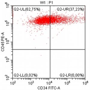
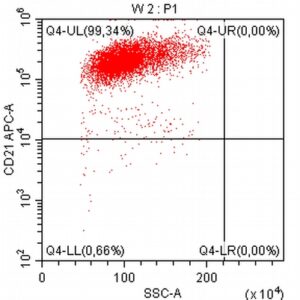
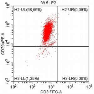
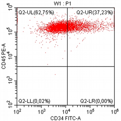
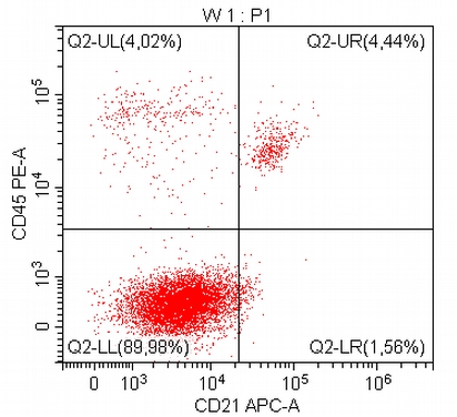
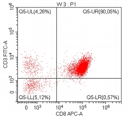
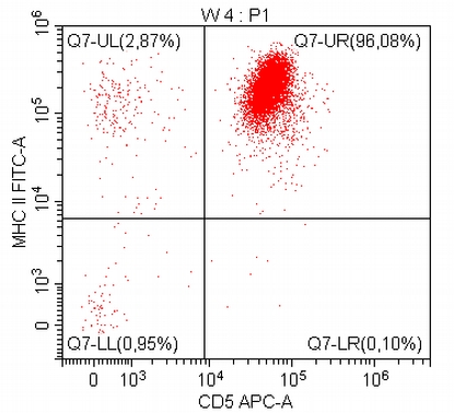
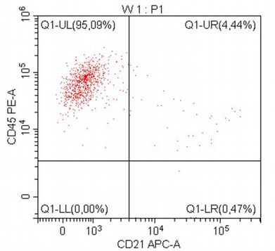
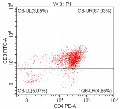
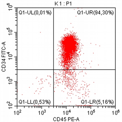
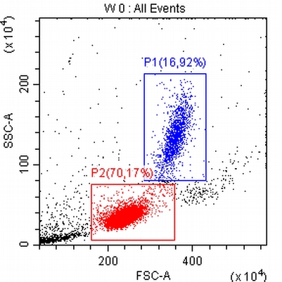
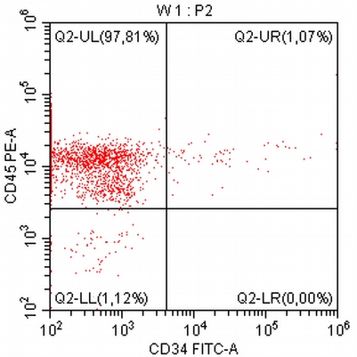
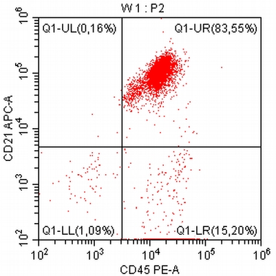
  • The results of the cytometric analysis showed that the dominant population in the examined lymph node consisted of medium to large cells with the immunophenotype CD45+ CD21+ CD79α+ MHC II+. This finding indicated a disruption of the physiological proportions between T and B lymphocytes in the lymph node and clearly suggested excessive proliferation of B lymphocytes.*

Taking into account the clinical data and the cytology results of the patient’s lymph nodes, immunophenotyping made it possible to resolve doubts about the final diagnosis — the presence of B-cell lymphoma was confirmed and appropriate therapy was initiated.

*Pawlak A, et al. Immunophenotypic characterization of canine malignant lymphoma: a retrospective study of cases diagnosed in Poland Lower Silesia, over the period 2011–2013. Veterinary and Comparative Oncology, 2014, 14, 52–59.


Shih Tzu with signs of generalized lymphadenopathy and marked lymphocytosis in peripheral blood

  • An 11-year-old female Shih Tzu with signs of generalized lymphadenopathy and marked lymphocytosis in peripheral blood persisting for about a year.
  • Histopathological examination of the excised submandibular lymph node performed at the beginning of the diagnostic process indicated reactive hyperplasia, which did not correlate with the clinical picture.
  • The immunophenotyping result indicated the presence of a clearly dominant population of cells in the examined lymph node with the immunophenotype CD45- CD3+ CD8+ CD5+ MHC II+. The presence of a large population of cells with the phenotype of cytotoxic T lymphocytes lacking expression of the lymphocyte marker (CD45) was indicative of their neoplastic transformation.

In this case, immunophenotyping enabled confirmation of a clinically significant lymphoma and, based on literature data, suggested its subtype as T-zone lymphoma (TZL).*

*Seelig DM, Avery P, et al. Canine T-zone lymphoma: unique immunophenotypic features, outcome, and population characteristics. J Vet Intern Med 2014; 28: 878-86.


Boxer with generalized lymphadenopathy and cytological suspicion of lymphoblastic lymphoma

  • A 7-year-old Boxer with generalized lymphadenopathy and cytological suspicion of lymphoblastic lymphoma
  • Cytometric analysis enabled the identification of a clearly dominant population of cells in the examined lymph nodes with the phenotype CD45+ CD3+ CD5+ CD4+, corresponding to helper T lymphocytes.Díky imunofenotypizaci byla přítomnost lymfomu rychle a jednoznačně potvrzena, navíc byl jeho původ stanoven na T buňky. According to the literature, the CD45+ CD4+ phenotype in T-cell lymphomas may predict an aggressive nature and rapid progression of the neoplastic disease, and Boxers are considered a breed with a high predisposition to this type of neoplasia*.**


*Avery PR, Burton JL, et al. Flow cytometric characterization and clinical outcome of CD4+ T-Cell Lymphoma in dogs: 67 cases. J Vet Intern Med 2014; 28: 538-546.
**Lurie DM, Milner RJ, et al. Immunophenotypic ad cytomorphologic subclassification of T-cell lymphoma in the boxer breed. Vet Immunology and Immunopathology 2008; 125: 102-110.


Mixed-breed dog in severe general condition with marked leukocytosis and the presence of atypical cells in a peripheral blood smear

  • A 9-year-old mixed-breed dog in severe general condition with marked leukocytosis, anemia, thrombocytopenia, and the presence of atypical cells in a peripheral blood smear
  • As a result of the blood cytometric analysis, a clearly dominant population of stem cells (CD34+) was identified, with concurrent expression of the B-lymphocyte marker (CD79α+).
    The phenotyped cells did not express CD14 or CD4, which made it highly probable to exclude a myelomonocytic hyperplasia lineage. In this case, immunophenotyping formed the basis for the final diagnosis of acute B-lymphoblastic leukemia (ALL-B)*.


* Vernau W, Moore PF. An immunophenotypic study of canine leukemias and preliminary assessment of clonality by polymerase chain reaction. Vet Immunol Immunopathol 1999; 69: 145-164.


Miniature Schnauzer showing no clinical symptoms with marked lymphocytosis persisting for several months

  • A 15-year-old Miniature Schnauzer showing no clinical symptoms, with marked lymphocytosis persisting for several months and no atypical cells present in peripheral blood
  • In the cytometric blood analysis, the dominant cell population displayed the phenotype CD45+ CD21+ CD79α+ MHC II+, corresponding to B lymphocytes. At the same time, the lack of expression of the stem cell marker (CD34) in the examined cells excluded the presence of acute lymphoblastic leukemia. In this case, the immunophenotyping result (in the context of the clinical data and the peripheral blood smear findings) made it possible to consider the presence of chronic B-cell lymphocytic leukemia (B-CLL) or, alternatively, small B-cell lymphoma (B-SLL) with blood involvement*.

*Comazzi S, Gelain ME, Martini V. Immunophenotype Predicts Survival Time in Dogs with Chronic Lymphocytic Leukemia. JVIM 2011: 25; 100-106.